HomeWithout LabelLeukoplakia And Erythroplakia Difference / red and white lesions of oral cavity - These investigators indicated that the erythroplakia is rare compared to oral leukoplakia.
Rabu, 03 Februari 2021
Leukoplakia And Erythroplakia Difference / red and white lesions of oral cavity - These investigators indicated that the erythroplakia is rare compared to oral leukoplakia.
Leukoplakia And Erythroplakia Difference / red and white lesions of oral cavity - These investigators indicated that the erythroplakia is rare compared to oral leukoplakia.. Other disease. simply put, if a white lesion in the oral cavity can be given a specific diagnosis it is not a. Leukoplakia is a white patch in the oral cavity.(medical term is leukoplakia). Leukoplakia or erythroplakia exhibiting moderate or severe dysplasia should be surgically removed if possible. Histologic appearance of leukoplakia indistinguishable from proliferative (verrucous). These differences are important, because they can impact a person's treatment options and prognosis (outlook).
Recognized potentially malignant disorders are leukoplakia, erythroplakia, palatal lesions in reverse smokers, submucous fibrosis, actinic cheilitis and lichen planus; Changes generally take place on your gums thickened or hardened areas. This difference becomes more pronounced with increasing age. It may also be linked to oral cancer and must be treated promptly. These investigators indicated that the erythroplakia is rare compared to oral leukoplakia.
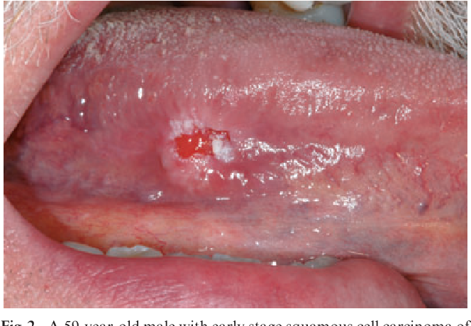
Figure 2 from Oral cancer and oral erythroplakia: an ... from ai2-s2-public.s3.amazonaws.com Leukoplakia exhibiting hyperkeratosis that is not frictional / reactive exhibits a malignant transformation rate of approximately 5%, similar to that of mild epithelial dysplasia. Erythroleukoplakia can therefore be considered a variant of either leukoplakia or erythroplakia since its appearance is midway between.20 erythroleukoplakia frequently occurs on the buccal mucosa in the commisural area (just. The prognosis and overall survival of a patient with oral cancer is dependent on the early detection of any lesion that might identify a patient with higher risk than normal or with early infiltration. The newest definitions for leukoplakia, erythroplakia, and smokeless tobacco keratosis are offered, along with a rationale for predicting malignant @article{bouquot1994oralla, title={oral leukoplakia and erythroplakia: Smoking is the most common cause. Cryotherapy and laser ablation have been used, although. The equivalent terms squamous intraepithelial neoplasia and oral intraepithelial neoplasia have not to date been universally accepted. I'm not a doctor but i don't think one is more prone to developing into cancer than the other.
A review and update.}, author={j.
Leukoplakia and erythroplakia are terms used to describe certain types of tissue changes that can be seen in the mouth or throat different cancers can start in each type of cell. The edges of the lesion are typically abrupt and the lesion changes with time. Erythroleukoplakia can therefore be considered a variant of either leukoplakia or erythroplakia since its appearance is midway between.20 erythroleukoplakia frequently occurs on the buccal mucosa in the commisural area (just. Erythroplakia is the clinical diagnostic term for a chronic red mucosal macule which cannot be given another specific diagnostic name, and cannot be attributed to traumatic, vascular or inflammatory causes, (i.e. When scraped, it bleeds easily. When leukoplakia speckled with red spots > erythroleukoplakia. (eds) premalignant conditions of the oral cavity. This difference becomes more pronounced with increasing age. The equivalent terms squamous intraepithelial neoplasia and oral intraepithelial neoplasia have not to date been universally accepted. Learn vocabulary, terms and more with flashcards, games and other study tools. Leukoplakia or erythroplakia exhibiting moderate or severe dysplasia should be surgically removed if possible. Learn how to identify and treat leukoplakia. Recognized potentially malignant disorders are leukoplakia, erythroplakia, palatal lesions in reverse smokers, submucous fibrosis, actinic cheilitis and lichen planus;
Leukoplakia is a firmly attached white patch on a mucous membrane which is associated with increased risk of cancer. Recognized potentially malignant disorders are leukoplakia, erythroplakia, palatal lesions in reverse smokers, submucous fibrosis, actinic cheilitis and 3 there are geographical differences with regard to gender distribution. Recognized potentially malignant disorders are leukoplakia, erythroplakia, palatal lesions in reverse smokers, submucous fibrosis, actinic cheilitis and lichen planus; The edges of the lesion are typically abrupt and the lesion changes with time. Both have high risk of malignant transformation 50%!!!
Medical problems 1 4 from image.slidesharecdn.com A review and update.}, author={j. Learn how to identify and treat leukoplakia. In most patients, the lesions. These white or red patches might be harmless. Start studying leukoplakia and erythroplakia. When leukoplakia speckled with red spots > erythroleukoplakia. Oral erythroplakia, of all precancerous oral lesions, carries the greatest threat of malignant transformation. In this video we will discuss the pathology of leukoplakia and erythroplakialeukoplakia and erythroplakia are premalignant conditions of oral cavity that.
For diagnosis and management coordinator:
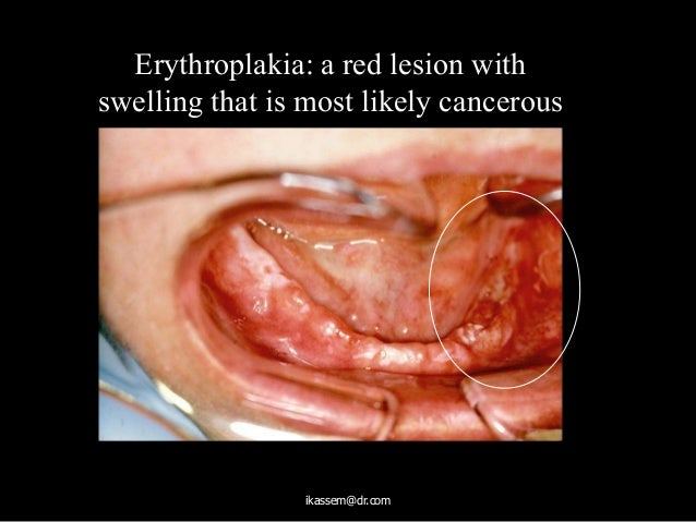
It is a health condition that in some patients, these may be red in appearance and referred to as erythroplakia, a condition that may result in cancer. But they can also be precancerous and contain abnormal cells. Erythroplakia is a red area in the mouth that bleeds easily. Brennan p., aldridge t., dwivedi r. Learn how to identify and treat leukoplakia. Erythroplakia is the clinical diagnostic term for a chronic red mucosal macule which cannot be given another specific diagnostic name, and cannot be attributed to traumatic, vascular or inflammatory causes, (i.e. Leukoplakias are usually diagnosed after the fourth decade of life and are six. Leukoplakia and erythroplakia are the two most common potentially malignant disorders of the oral cavity. Oral erythroplakia, of all precancerous oral lesions, carries the greatest threat of malignant transformation. Most cases of leukoplakia and erythroplakia are seen in adults older than 50 years who have risk factors (discussed in etiology). Oral leukoplakia is defined as. I'm not a doctor but i don't think one is more prone to developing into cancer than the other. Leukoplakia appears as thickened, white patches on your gums, cheeks and bottom of your mouth leukoplakia usually occurs on your gums, the insides of your cheeks, the bottom of your mouth along with raised, red lesions (speckled leukoplakia or erythroplakia), which are more likely to show.
Other disease. simply put, if a white lesion in the oral cavity can be given a specific diagnosis it is not a. This is a red patch that can be both flat or slightly raised. Possible oral lesions include erythroplakia, leukoplakia, or erythroleukoplakia. Oral hairy leukoplakia in immunocompromised individuals. The prognosis and overall survival of a patient with oral cancer is dependent on the early detection of any lesion that might identify a patient with higher risk than normal or with early infiltration.
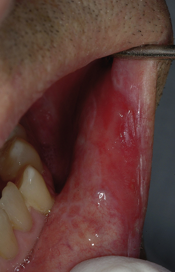
27: Erythroplakia (erythroplasia) | Pocket Dentistry from pocketdentistry.com I'm not a doctor but i don't think one is more prone to developing into cancer than the other. Recognized potentially malignant disorders are leukoplakia, erythroplakia, palatal lesions in reverse smokers, submucous fibrosis, actinic cheilitis and lichen planus; Erythroplakia and speckled leukoplakia are more likely to exhibit dysplasia or carcinoma upon microscopic examination. The prognosis and overall survival of a patient with oral cancer is dependent on the early detection of any lesion that might identify a patient with higher risk than normal or with early infiltration. Erythroplakia is a red area in the mouth that bleeds easily. Leukoplakia is a white patch in the mouth. (eds) premalignant conditions of the oral cavity. Brennan p., aldridge t., dwivedi r.
Erythroleukoplakia can therefore be considered a variant of either leukoplakia or erythroplakia since its appearance is midway between.20 erythroleukoplakia frequently occurs on the buccal mucosa in the commisural area (just.
Leukoplakia is a white patch in the oral cavity.(medical term is leukoplakia). Changes generally take place on your gums thickened or hardened areas. The equivalent terms squamous intraepithelial neoplasia and oral intraepithelial neoplasia have not to date been universally accepted. Histologic appearance of leukoplakia indistinguishable from proliferative (verrucous). The newest definitions for leukoplakia, erythroplakia, and smokeless tobacco keratosis are offered, along with a rationale for predicting malignant @article{bouquot1994oralla, title={oral leukoplakia and erythroplakia: But they can also be precancerous and contain abnormal cells. It's natural to feel overwhelmed, but we're here to guide you through the process. Leukoplakia and erythroplakia are the two most common potentially malignant disorders of the oral cavity. Leukoplakia and erythroplakia are terms used to describe certain types of tissue changes that can be seen in the mouth or throat different cancers can start in each type of cell. The newest definitions for leukoplakia, erythroplakia, and smokeless tobacco keratosis are offered, along with a rationale for predicting malignant transformation and for treatment planning of these most important precancers. Start studying leukoplakia and erythroplakia. In this video we will discuss the pathology of leukoplakia and erythroplakialeukoplakia and erythroplakia are premalignant conditions of oral cavity that. The term oral dysplasia is synonymous with intraepithelial neoplasia as used when discussing the cervix.
The prognosis and overall survival of a patient with oral cancer is dependent on the early detection of any lesion that might identify a patient with higher risk than normal or with early infiltration leukoplakia and erythroplakia. Leukoplakias are usually diagnosed after the fourth decade of life and are six.